Nikon A1R MP+ Multiphoton Upright Microscope
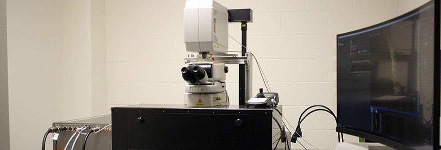
The Nikon A1R MP upright multiphoton microscope can be used to capture fluorescent images deep into tissues. Cells and live organs can be imaged at a greater depth than is currently possible with typical laser-scanning confocals. Non-intravital applications for multiphoton microscopy include tracking fluorescent protein-expressing immune cells and visualizing the dynamics of drug- and metal-containing nanoparticles.
Housed within a dark enclosure, a height-adjustable motorized XY stage can accommodate multiple model organisms and live animals, in addition to cultured cells. The tunable long wavelength IR laser allows for simultaneous imaging of a variety of fluorescent probes.
Nikon A1R MP+ Multiphoton Brochure
Multiphoton system for intravital imaging
Key Features
- The A1R MP integrates a four-channel GaAsP (gallium arsenide phosphide) detector unit to allow simultaneous imaging of up to four probes.
- In addition, a fully tunable A1-DUVB-2 GaAsP spectral detector allows for user definable emission bandwidths instead of reliance on fixed emission filters. The GaAsP detector is more sensitive than conventional PMTs and is useful with photon limited samples.
- A LU-N4 4-laser unit is equipped with 405, 488, 561, and 640nm lasers.
- A Chameleon Discovery femtosecond pulsed laser capable of tunable output of 700-1300nm with auto alignment.
- A Resonant scanner capable of speeds of 30 fps (512x512) and 420 fps (512x32).
- A Galvano scanner with speed acquisitions capable of 10 fps (512x512) and 130 fps (512x32), high-resolution of imaging up to 4096x4096 pixels.
- Nikon Apochromat LWD 25X lens (NA 1.10W water dipping, WD 2.0mm objective).
-
A specialized objective optimized for imaging cleared tissues: CFI90, LWD 20X (NA 1.0 GLYC, WD 8.2 mm).
- High speed Piezo drive for Z-stacking and large tiled images.
- Nikon Elements Software
Gallery
In vivo microscopy scan of a venule and capillaries in the mouse skeletal muscle with plasma tracer (green; FITC-dextran) and leukocytes (yellow; stained with Rhodamine 6G) interacting with venular endothelium. Scan collected with Nikon A1R MP+ Multiphoton Microscope by Stephanie Milkovich, Paulina Kowalewska and Chad Steele.


